Below are speaker bios and abstracts for MagCAN 2023. Click below for more details on a group or speaker.
All times are central time.
Visit the conference webpage for the full conference agenda and other details.
Group 1 (August 24) - MicroMagnetic Neurostimulation, Moderator: Shelley Fried (Harvard)
1:30-2:00pm - Renata Saha, UMN
Exploring Micromagnetic Stimulation as An Alternative to Electrode Stimulation: Prospects & Challenges
Abstract
Micromagnetic stimulation (μMS), although still in its infancy, is a neuromodulation technique where the devices are deep brain stimulation (DBS)-like implants, but they operate on the physics of transcranial magnetic stimulation (TMS). Essentially, they are micrometer-sized coils or microcoils (μcoils) which when driven by an alternating current, generates a time varying magnetic field. As per Faraday’s Laws of Electromagnetic Induction, this magnetic field induces an electric field that activates the neurons. Spatially selective and galvanic contact-free neural activation coupled with MRI-safe implant performance are some of the unique features of µMS that make this neuromodulation technique so unique. The only drawback of this technology is that the µcoils need to drive a high amplitude current waveform for successful neural activation.
In this talk, we will discuss the designing, fabrication and testing of two micromagnetic implants – the MagneticPen (MagPen), a solenoid-shaped single µcoil prototype and the Magnetic Patch (MagPatch), a rectangular helix shaped planar µcoil array prototype. The efficacy of micromagnetic activation using MagPen has been tested over rat hippocampal CA3-CA1 synaptic pathway in vitro, on the medial forebrain bundle (MFB) of rodents for the study of striatal dopamine release in vivo and the vagus nerve to demonstrate fiber-specific activation of the nerve in vivo. The experimental testing on rodents confirmed the already existing unique features of µMS in addition to contributing several unique features to this neuromodulation technique. For instance, fiber-specific activation, the importance of the directionality of induced electric field on successful neural activation and several others. The MagPatch array had been designed to offer neural activation at the single cell resolution and study the effect of the directionality of induced electric field from each of the µcoils in the array for successful neuron activation. Finally, using the MagPen prototype we have been able to demonstrate the dose-response relationship for µMS on the rat sciatic nerve. The impact of this dose-response curve lies on the fact that it addresses the controversy in this research community whether the stimulation from these μcoils is a thermal effect or an effect from micromagnetic stimulation. In summary, we will explore the efficacy of µMS in several experimental settings, both in vitro and in vivo, making some addition to the unique features of µMS in the process. In addition, we will strategize techniques using soft magnetic material (SMM) cores at the center of these µcoils to reduce their high-power thresholds in µMS.
Biography

Dr. Renata Saha just successfully defended her PhD at the Department of Electrical & Computer Engineering at the University of Minnesota, Twin Cities. She received her Bachelor in Technology (B.Tech) from National Institute of Technology, Durgapur, India. In the Summer of 2017, she was an undergraduate research intern at Conseil européen pour la Recherche Nucléaire (CERN), Geneva, Switzerland. She is the recipient of 3-year College of Science & Engineering Fellowship from the University of Minnesota, Women in Technology Scholarship 2021-2022 from Cadence Design Systems, elected iREDEFINE Fellow 2022 by Electrical & Computer Engineering Department Heads Association (ECEDHA) and recipient of Graduate Neuromodulation Research Fellowship 2022-2023 from Minnesota’s Discovery, Research and InnoVation Economy (MnDRIVE). She has published over 27 journal articles, 3 US patents and 2 book chapters. She is the recipient of 3 Best Poster Awards and 2 Travel Awards. Her research interest includes designing and development of microelectromechanical systems (MEMs)-based micromagnetic devices for their application in healthcare.
2:00-2:30pm - Giorgio Bonmassar, Harvard
Short-pulsed micro-magnetic stimulation of the vagus nerve
Abstract
Vagus Nerve Stimulation (VNS), commonly used for drug-resistant epilepsy and depression treatment. VNS involves stimulating specific vagal fibers, but risks stimulating cardiac fibers, causing rare but serious bradyarrhythmia side effects. This risk is hard to mitigate due to VNS's electrode-based technology, which indiscriminately activated fibers. Micro-magnetic stimulation (μMS), is introduced, aiming for focused vagus nerve stimulation using implanted micro-coils in rats, for a more targeted fiber stimulation. Comparing with electrical VNS, μM-VNS reduces respiration rate temporarily without affecting heart rate (HR), suggesting potential heart-related side effect reduction. Numerical simulations aided optimal coil placement, estimating the induced electric field distribution. Transmission emission microscope images provided very high-resolution images of the cervical vagus nerve in rats, which revealed two different populations of nerve fibers categorized as large and small myelinated fibers and identified clusters of large fibers ideal for μMS targeting. I will be presenting latest multichannel Figure-8 micro-coils for μM-VNS. This research explores μMS as an alternative to VNS with potential for fewer heart-related risks.
Biography
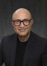
2:30-3:00pm - Jae-lk Lee, Harvard
Magnetic Intracochlear Stimulation: A Pilot Study
Abstract
Cochlear implants (CIs) are auditory prostheses that electrically stimulate the auditory nerve to partially restore hearing in people with severe to profound hearing loss. The inability to create narrow regions of spectral activation is thought to limit the performance of existing CIs. Previous studies, outside the cochlea, have shown that magnetic stimulation from small, implantable coils (micro-coils) confines activation narrowly, raising the possibility that coil-based stimulation of the cochlea could provide improved spectral resolution. This study explored the potential of magnetic micro-coil stimulation in the cochlea to improve spectral resolution of CIs. Magnetic stimulation from micro-coils was delivered to multiple locations in the mouse cochlea, and the spread of activation was measured using a multielectrode array inserted into the inferior colliculus. Responses to magnetic stimulation were compared to analogous experiments with conventional microelectrodes as well as to responses to auditory monotones. The extent of activation with micro-coils was found to be approximately 60% narrower compared to electric stimulation and similar to the spread arising from acoustic stimulation. The dynamic range of coils was also found to be more than three times larger than that of electrodes, further supporting a smaller spread of activation. The narrower spread of activation with micro-coils suggests that a larger number of independent spectral channels can be created, which may improve auditory outcomes.
Biography

My primary research focus is on the development of more effective and innovative techniques to artificially stimulate neurons for prosthetic devices. Throughout my research, I have delved into the responses of retinal neurons to electric stimulation, aiming better replication of physiological signaling patterns. In addition to my work on retinal neurons, I have recently been investigating the feasibility of employing magnetic stimulation as an alternative to electric stimulation in cochlear implants. Encouragingly, preliminary data indicates that magnetic coils can robustly stimulate the auditory pathway, surpassing conventional electrodes by offering improved spectral resolution. This exciting discovery suggests the potential to significantly enhance clinical outcomes for individuals relying on cochlear implants.
3:00-3:30pm - Hui Ye, Loyola U. Chicago
Cellular mechanisms underlying state-dependent neural inhibition with magnetic stimulation
Abstract
Novel stimulation protocols for neuromodulation with magnetic fields are explored in clinical and laboratory settings. Recent evidence suggests that the activation state of the nervous system plays a significant role in the outcome of magnetic stimulation, but the underlying cellular and molecular mechanisms of state-dependency have not been completely investigated. We recently reported that high frequency magnetic stimulation could inhibit neural activity when the neuron was in a low active state. In this work, we investigate state-dependent neural modulation by applying a magnetic field to single neurons, using the novel micro-coil technology. High frequency magnetic stimulation suppressed single neuron activity in a state-dependent manner. It inhibited neurons in slow-firing states, but spared neurons from fast-firing states, when the same magnetic stimuli were applied. Using a multi-compartment NEURON model, we found that dynamics of voltage-dependent sodium and potassium channels were significantly altered by the magnetic stimulation in the slow-firing neurons, but not in the fast-firing neurons. Variability in neural activity should be monitored and explored to optimize the outcome of magnetic stimulation in basic laboratory research and clinical practice. If selective stimulation can be programmed to match the appropriate neural state, prosthetic implants and brain-machine interfaces can be designed based on these concepts to achieve optimal results.
Biography

Dr. Hui Ye received his PhD degree in Biomedical Engineering from Case Western Reserve University in Cleveland, OH. His thesis work investigated the neural control and biomechanics underlying adaptive behavior in animals. He received postdoctoral training from Toronto Western Research Institute and University of Toronto, and was awarded a postdoctoral fellowship from the Heart and Stroke foundation of Canada. Ye joined Loyola University Chicago as an assistant professor in 2012, and was promoted to associated professor in 2018.
Dr. Ye is currently working on a NIH-support project to investigate the cellular and molecular mechanisms underlying electric and magnetic stimulation. He has published more than 40 papers in Neuroscience, Biophysics, and Biomedical Engineering. He teaches Loyola’s Neuroscience lab courses that include neurophysiology and computational neuroscience methods.
Group 2 (August 24) - Magnetic Neuron Sensing, Moderator: William Rippard (NIST)
3:45-4:15pm - Lawrence L Wald, Harvard
Magnetic Particle Imaging (MPI) for functional brain imaging; rodent and human-scale devices
Abstract
Biography
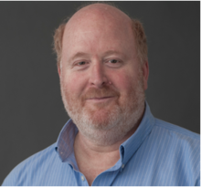
Lawrence L. Wald, Ph.D., is currently a Professor of Radiology at Harvard Medical School and Affiliated Faculty of the Harvard-MIT Division of Health Sciences Technology. He received a Ph.D. in Physics from the University of California at Berkeley in 1992 under the direction of Prof. E.L. Hahn with a thesis related to the optical detection of NMR. He obtained further (postdoctoral) training in Physics at Berkeley and Radiology and MRI at the University of California at San Francisco (UCSF). He began his academic career as an Instructor at the Harvard Medical School at McLean Hospital. Since 1998 he has been at the Massachusetts General Hospital Dept. of Radiology in the NMR Center (now the A.A. Martinos Center for Biomedical Imaging). His work has explored the benefits and challenges of highly parallel MRI and its application to highly accelerated image encoding and parallel excitation and ultra-high field MRI (7 Tesla) methodology for brain imaging. This has included improved methods for RF coil arrays, parallel transmit methods, acquisition sequences, matrix shimming, peripheral nerve modeling, and gradient coil design. His lab also focuses on motion mitigation methods and portable MRI technology and is developing a prototype Magnetic Particle Imaging scanner for human functional brain imaging.
4:15-4:45pm - Samuel Oberdick, NIST
Emerging Applications of Magnetic Nanoparticles for MRI Contrast: Positive T1 Contrast at Low Field and “Color” Contrast at High Field
Abstract
Biography

Dr. Samuel Oberdick received his PhD from Carnegie Mellon University in physics in 2016, where he studied magnetic nanomaterials and devices. From 2017 to 2019 he was a National Research Council postdoctoral fellow working with the Magnetic Imaging Group in the Applied Physics Division at NIST, Boulder. He is currently a research associate with the University of Colorado, Boulder Physics Department and is also an associate of NIST, Boulder. His present research is focused on the intersection of magnetic nanomaterials and biological systems. Specifically, he is interested in using magnetic nanomaterials for biosensing applications and development of contrast agents for magnetic resonance imaging.
4:45-5:15pm- Aviad Hai UW-Madison
Wireless Bioelectromagnetic Brain Sensors
Abstract
There is currently a concerted effort to develop the necessary technologies to record and stimulate neural activity across the entire volume of the mammalian brain. Recent engineering advancements have propelled electrode- and optical-based devices, achieving nanometer scale spatial resolution and impressive signal-to-noise ratio and temporal response. However, these probes usually require a tethered connection and provide access to relatively small areas in the nervous system. Developing modalities for whole-brain direct recording and stimulation of neural signals, will allow neuroscientists and neurologists to study and treat the brain network directly and as a whole, and will surely elevate brain science and medicine to new heights. I will describe the development of wireless, implantable electronic probes that are able to transduce electromagnetic fields in the brain, as well as the application of molecular agents for large volume in vivo measurements of neurotransmitter dynamics in live mammals. These strategies pave the way towards functional studies of neural activity across wide brain regions with molecular and electrophysiological specificity.
Biography
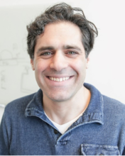
Dr. Aviad Hai is a neuroengineer and a neuroscientist whose group focuses on developing implantable and injectable agents for accessing the nervous system—towards achieving a broader understanding of brain function. Dr. Hai is the recipient of the 2020 NIH Director’s New Innovator award, the NIH NIBIB Mentored Research Scientist Development Award (K01), a long-term fellowship from the European Molecular Biology Organization (EMBO) and a fellowship from the Edmond & Lily Safra Center for Brain Sciences. In 2019, Dr. Hai joined the Department of Biomedical Engineering at the University of Wisconsin-Madison as an assistant professor, where his group is developing cutting edge electrical, magnetic, and electromagnetic sensors for large-scale recording and stimulation of brain activity. Dr. Hai received his Ph.D. from the Hebrew University of Jerusalem under the guidance of Prof. Micha Spira (Neurobiology) and Prof. Joseph Shappir (Electrical Engineering), where he pioneered nano-scale devices for multiplexed recording of neuronal intracellular signals. He later joined Prof. Alan Jasanoff at the department of Biological Engineering at the Massachusetts Institute of Technology, where he developed wireless implantable sensors that detect electromagnetic fields in the brain, and molecular technologies for direct imaging of neurotransmitter dynamics across large volumes of the brain. His efforts cohere with the goal of providing neuroscientists and neurologists with a complete toolbox for understanding and modulating electrical and neurochemical activity across the entire volume of the brain in parallel.
Group 3 (August 25) - Magnetic Neurostimulation, Moderator: Andrew Whalen (Yale)
9:00-9:30am - Alex Opitz, UMN
Transcranial magnetic stimulation in animal models and humans – bridging scales with computational models
Abstract
Transcranial magnetic stimulation (TMS) is a non-invasive brain stimulation technique which modulates cortical excitability beyond the stimulation period. Based on the physical principle of electromagnetic induction, TMS is capable of depolarizing cortical neurons through the intact scalp and skull of awake, non-anesthetized subjects. Despite its clinical use (e.g., treatment of pharmacoresistant depression) it remains unclear how TMS-mediated changes in synaptic transmission, neuronal excitability, and network activity exert beneficial effects in some healthy subjects and patients. Animal models are crucial to advance our understanding of TMS mechanisms as they allow invasive recordings not available in humans. However, in order to translate findings from animals to humans, great care has to be taken. Due to differences in brain/head anatomy, TMS induced electric fields will differ across species. Similarly, neuron morphology, biophysics, and alignment with respect to the TMS electric field can lead to different stimulation effects. This can lead to findings from basic animal studies that are difficult to translate to human TMS applications. I will discuss challenges of identifying suitable TMS parameters in animal models and highlight the utility of multi-scale computational models in these efforts.
Biography

Alexander Opitz is an Associate Professor in the Department of Biomedical Engineering at the University of Minnesota. His research focuses on developing non-invasive brain stimulation technologies. Dr. Opitz has a particular interest in the underlying biophysics and physiology of transcranial magnetic stimulation (TMS) and transcranial electric stimulation (TES). He believes it is necessary to study the effect of brain stimulation across various levels of investigation to make progress. Thus, research in his lab spans from computational modeling of electric fields and their effect on neurons, to electrophysiological recordings of brain activity in animal models and humans. His lab is further developing closed-loop stimulation technologies to improve the effectiveness of TMS.
9:30-10:00am - Svenja Knappe, UC-Boulder
Non-invasive functional brain imaging with an array of small uncooled quantum magnetometers
Abstract
Biography

10:00-10:30am - Padmavathi Sundaram, Harvard
Cellular Mechanisms of Transcranial Magnetic Stimulation in the Cerebellum
Abstract
In this talk, I will describe how my lab is developing a cellular level understanding of how TMS activates neurons in the cerebellum of humans. For this we use electrophysiology experiments in an in vitro cerebellum model and computational modeling the induced electric fields in the tissue. Since the local anatomy and electrophysiology of the cerebellum are evolutionarily conserved, the fundamental neural dynamics will generalize. The combination of invasive electrophysiology in the in vitro cerebellum, the in vivo human TMS studies combined with electric field simulations will likely allow better understanding of the cellular basis/biophysics of TMS, opening new applications of TMS for studying the role of the cerebellum in human brain function.
Biography

Padmavathi Sundaram is an Assistant Professor at the Martinos Center for Biomedical Imaging at Massachusetts General Hospital/Harvard Medical School. She received her PhD in Electrical Engineering from Stanford University and did her postdoctoral work at Brigham and Women’s Hospital and Boston Children’s Hospital. Her research is focused on bridging electrophysiology, brain stimulation, computational modeling and neuroimaging. On the neuroscience side, her lab is interested in using these different approaches to record from, stimulate the cerebellum in both humans and in vitro animal models.
10:30-11:00am - Andrew Whalen, Yale
Thermal safety considerations for implantable micro-coil design
Abstract
Micro magnetic stimulation of the brain via implantable micro-coils is a promising novel technology for neuromodulation. These implants can require high levels of current flow during operation; therefore, their operational thermodynamic profile must be considered to ensure such devices are safe for long-term implantation. ISO standard 14708-1 designates a thermal safety limit of 2°C temperature increase for active implantable medical devices.
Using bent wire micro-coils submerged in a water bath, we measured the thermal gradients generated near the surface of the micro-coils during stimulation for a range of stimulation conditions consistent with prosthetic device operation. I will talk about how careful selection of stimulation parameters (voltage amplitude, stimulus frequency, repetition rate) and coil construction materials (insulation, coil conductor material) can achieve 5-6 fold decrease in the thermal impact of coil stimulation to limit temperature increases to ensure the thermal gradients generated remained below the 2°C safety limit for all stimulation parameters tested.
Biography

Andrew J Whalen received his undergraduate and doctoral degrees in mechanical engineering from the Pennsylvania State University in 2008 and 2017, with a research focus on fusing control engineering with the physiology of fundamental neuronal mechanisms to develop better treatment methods for control of brain pathophysiology. He joined Dr. Shelley Fried’s lab in 2019 as a postdoctoral research fellow at Massachusetts General Hospital and Harvard Medical School, studying retinal prosthetic neuromodulation and micro-magnetic stimulation in small animal models. He is now an Associate Research Scientist in the Department of Neurosurgery at Yale University working in collaboration with Dr. Steven Schiff.
His research interests broadly include many facets of neuroscience, from computational modeling of complex nonlinear brain networks, to modeling and control of infectious disease, measurement and stimulation of neural tissues, and dynamical mechanisms underpinning neurological diseases.
11:00-11:30am - Ravi Hadimani, VCU
Transcranial Magnetic Stimulation: A non-invasive neuromodulation technique for altering neuronal networks in the brain
Abstract
Transcranial Magnetic Stimulation (TMS) can tune brain functions non-invasively, safely, and effectively without the need for surgery or drugs. Thus, it can enable the treatment of several debilitating neurological obsessive-compulsive disorders, and migraine [1]. My lab has designed and fabricated an anatomically accurate human brain phantom that can be used to test the feasibility and safety of several TMS protocols. We have investigated a feasibility study of combined TMS and DBS using brain phantom in collaboration with the VCU Dept. of Neurosurgery [2], [3]. We have also designed and fabricated novel focal stimulation coils based on novel soft ferromagnetic materials that can stimulate only a local region of the primary motor cortex. We are currently working to experimentally verify the results from coil design in rats in collaboration with the Dept. of Neurology at VCU [4]. We are also working to establish an accurate mechanism underlying TMS by investigating the neuronal firing patterns in several brain regions induced by cortical stimulation and by establishing the role of individual nuclei in affecting other nuclei of the motor circuitry. My team has also designed a TMS coil configuration that can stimulate multiple sites simultaneously and vary sites of stimulation without moving the coils physically. These new TMS techniques will enable the future development of effective TMS protocols for the diagnosis and treatment of several neurological and psychiatric disorders.
References
- S. Rossi et al., “Safety and recommendations for TMS use in healthy subjects and patient populations, with updates on training, ethical and regulatory issues: Expert Guidelines,” Clin. Neurophysiol., 132, p. 269, 2021.
- H. Magsood, et al., “Safety Study of Combination Treatment: Deep Brain Stimulation and Transcranial Magnetic Stimulation,” Front. Hum. Neurosci., 14, p. 123, 2023.
- H. Magsood and R. L. Hadimani, “Development of anatomically accurate brain phantom for experimental validation of stimulation strengths during TMS,” Mater. Sci. Eng. C, 120, p. 111705, 2021.
- I. C. Carmona, D. Kumbhare, M. S. Baron, and R. L. Hadimani, “Quintuple AISI 1010 carbon steel core coil for highly focused transcranial magnetic stimulation in small animals,” AIP Adv., 11, p. 025210, 2021.
Biography

Dr. Hadimani an Associate Professor and the Director of the Biomagnetics Laboratory at the Department of Mechanical and Nuclear Engineering of Virginia Commonwealth University. He is currently on sabbatical as a Visiting Associate Professor at Harvard Medical School, Harvard University. He has founded the IEEE Joint Magnetics and Engineering in Medicine and Biology Society’s Richmond Chapter, and he is the current vice chair of the chapter. He is an Associate Editor of the journals, Frontiers of Neuroscience and American Institute of Physics (AIP) Advances.
Dr. Hadimani’s research focuses on biomagnetic materials and devices for biomedical applications, magnetocaloric refrigeration, and energy harvesting. He has developed a first-of-a-kind anatomically accurate brain phantom for validating neuromodulation procedures that are commercialized through the university spin-off company RAM Phantoms LLC. Dr. Hadimani has received several international awards, including the UK Energy Innovation Award the International Young Scientist Fellowship by the National Natural Science Foundation of China (NSFC). He also received the Engineer of the Year award from the Richmond Joint Engineers’ Council in 2021. He has authored more than 100 peer-reviewed original research papers, more than 210 international conference papers, 12 current and pending patents, several invited trade magazine articles, a book, and 3 book chapters to date.
Dr. Hadimani has a ‘first class’ honors degree in Mechanical Engineering from Kuvempu University, India, an MS in Mechatronics from the University of Newcastle, UK, and a Ph.D. in Electrical Engineering from Cardiff University, UK. He has served as a Project Scientist at the Institute of Materials Research and Innovation of the University of Bolton, UK. He was an Adjunct Assistant Professor and Associate Scientist at Iowa State University and was also an Associate of Ames Laboratory, US Dept. of Energy.
Group 4 (August 25) - Brain & Brain-inspired Technologies, Moderator: Ravi L Hadimani (VCU)
1:30-2:00pm - Kendall Lee, Mayo
Next Generation Smart DBS and Neuromodulation
Abstract
Our team has pioneered real-time imaging of the response of the brain during DBS in both humans and animals. Using BOLD-fMRI imaging techniques, we are identifying brain regions and networks that are activated by DBS and are studying how patterns of DBS-invoked brain activation change after long periods of stimulation. Although its mechanism of action is poorly understood, we know that DBS causes the release of chemicals called “neurotransmitters” in specific brain regions, depending on the site of stimulation, and we believe that stimulation-induced neurotransmitter release plays a critical role in the therapeutic benefits of DBS. The success of DBS depends on the amplitude and frequency parameters of stimulation. Today’s DBS systems deliver continuous stimulation at pre-set parameters in an open-loop mode without regard to potential changes in neural activity or patient behavior. We believe that tracking the dynamics (pattern and amount) of neurotransmitter release could serve as a feedback signal to develop a closed-loop smart system of DBS in which stimulation parameters are varied dynamically to optimize neurotransmitter release and maximize therapeutic effects for the patient. Our research and engineering team at Mayo has developed a neurotransmitter sensing system called WINCS (wireless instantaneous neurotransmitter concentration sensor), which is a device able to monitor stimulation-induced neurotransmitter release. We are now working to create and test WINCS MAVEN, the first integrated closed-loop DBS system for human use. Our clinical team was among the first to show neuromodulation therapy to be a viable treatment option for obsessive compulsive disorder and Tourette’s syndrome. We are now working to expand the application of DBS to other neurologic and psychiatric disorders including addiction.
Biography

2:00-2:30pm - William (Bill) Rippard, NIST
Single Flux Quantum Based Neuromorphic Computing
Abstract
Neuromorphic computing promises to dramatically improve the efficiency of certain computational tasks, such as perception and decision making. While software and specialized hardware implementations of neural networks have made tremendous progress, both implementations are still many orders of magnitude less energy efficient than the human brain. I will discuss how our new superconducting platform, based on dynamically reconfigurable magnetic Josephson junctions with spiking energies less than one attojoule, could lead to large-scale neuromorphic systems with better energy efficiency than the human brain and present experimental measurements of circuits based on them.
Biography

William H. Rippard is the Group Leader of the Spin Electronics Group in the Quantum Electrodynamics Division. The Group develops and advances metrologies for spin-based devices focusing on transport of electrons, heat flow, and spin excitations for advancing novel computing devices, neuromorphic hardware, and quantum-enhanced magnetic sensing. Their measurements concentrate on high-frequency behaviors, properties, and imaging of nanoscale devices and hybrid (e.g. superconducting/ferromagnet and CMOS/ferromagnet) structures for advanced computing. Dr Rippard received BS degrees in both Physics and Mathematics from the University of Florida in 1994 and a PhD in Applied Physics from Cornell University in 2000. Bill joined NIST as an NRC postdoctoral in 2000 fellow and became a permanent staff member in 2004 pursing metrology in spin-based devices with the goal of better understanding and utilizing the spin of the electron for practical applications in nanoscale devices. He has over 70 peer reviewed publications including high-profile journals such as Nature and Physical Review Letters. He received the Presidential Early Career Award for Scientists and Engineers (PECASE) in 2008 along with Silver and Bronze awards during his time at NIST along with a patent in 2019. He served as Acting Division Chief for the Quantum Electromagnetics Division for the last two years before stepping down in March 2023.
2:30-3:00pm - Chris Rourk, Jackson Walker
Ferritin-assisted deep brain stimulation
Abstract
Ferritin has electrical properties that make it useful both in engineered electronic components and intriguing with its presence in the mammalian brain. A novel function of biological ferritin has been proposed for neural processing in catecholaminergic neurons. The observed concentrations of ferritin and neuromelanin in those neurons may allow electron tunneling or similar functions to be used in neural processing. Electron tunneling behavior can be observed over distances as great as 12 nm through individual ferritin particles (1). Such tunneling may be magnon-assisted, resulting from the spintronic and magnetic properties of cellular ferritin. Electron tunneling can also be induced in fixed human SNc tissue (2) and there is evidence of tunneling associated with cellular ferritin in the retina, the cochlea, macrophages, mitochondria and other tissues (3). This diversity suggests that ferritin-mediated tunneling may be a widely-occurring phenomenon in cellular tissues and could provide a function for otherwise inexplicable presence of ferritin in vivo. Long-distance tunneling, over distances of 80 microns, has been observed in ferritin structures similar to those found in these tissues (4). Together these findings are consistent with the hypothesis that neuronal ferritin may modulate or mediate novel forms of neural processing in vivo. If so, there is a potential to better understand this novel function in neuronal circuits and explore the potential for developing ferritin-optimized therapies and diagnostics, including deep brain stimulation.
Rourk, Christopher J. Ferritin and neuromelanin “quantum dot” array structures in dopamine neurons of the substantia nigra pars compacta and norepinephrine neurons of the locus coeruleus." Biosystems 171 (2018): 48-58
Rourk,Christopher J. Indication of quantum mechanical electron transport in human substantia nigra tissue from conductive atomic force microscopy analysis. Biosystems 179 (2019): 30-38
Perez, Ismael Diez, et al. Electron tunneling in ferritin and associated biosystems. IEEE Transactions on Molecular, Biological and Multi-Scale Communications (2023).
Rourk, Christopher, et al. Indication of Strongly Correlated Electron Transport and Mott Insulator in Disordered Multilayer Ferritin Structures (DMFS). Materials 14.16 (2021): 4527.
Biography

Christopher (Chris) Rourk received a B.S.E.E. cum laude in 1985 from University of Florida, an M. Eng. In Electric Power Engineering from Rensselaer Polytechnic Institute in 1987 and a Juris Doctor cum laude from Georgetown University Law Center in 1995. He is a partner in the intellectual property department at Jackson Walker LLP in Dallas, TX and a former research scientist for Westinghouse Electric Corporation in the area of electric and magnetic field analysis of rotating machinery, and for the U.S. Nuclear Regulatory Commission on topics including the effect of lightning on nuclear power plants. He has been independently investigating electron tunneling associated with ferritin in biological systems since 2017.
3:00-3:30pm - Brandon Zink, UMN
Implementation of neuromorphic computing tasks with stochastic computing in computational random-access memory (SC-CRAM)
Abstract
Neuromorphic computing takes inspiration from biological nervous systems to improve energy efficiency when processing large data sets or performing brain-inspired tasks such as recognition or classification. Stochastic computing is a promising solution for neuromorphic systems since it benefits from high noise resiliency and can perform complex arithmetic functions with a small number of logic gates. The main shortcoming of stochastic computing is the large number of transistors required to generate random bit-streams using CMOS based technologies thus drastically increasing the total computation delay and energy consumption. In this work, we present a solution where stochastic computation and stochastic bit stream generation are performed in parallel using a spintronic based in-memory computing architecture called computational random access memory, which we called SC-CRAM. We demonstrate that SC-CRAM can efficiently perform arithmetic functions key for neuromorphic computing such as hyperbolic tangent and multiplication. By embedding the processing and stochastic bit-stream generation within the same CRAM cells, the advantages of stochastic computing can be leveraged without the additional costs of random number generation with CMOS platforms.
Biography
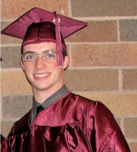
I am a Postdoctoral researcher for the Electrical Engineering department at the University of Minnesota. I hold BS and MS degrees in Physics from the University of Wisconsin – La Crosse and the University of Minnesota Duluth, respectively, and I recently received a PhD in Electrical Engineering from the University of Minnesota Twin Cities. I currently work with the Nanospin research group at the University of Minnesota, led by professor Jian-Ping Wang. My research involves fabrication and experimentation on magnetic tunnel junctions to examine their prospects in next generation, non-Boolean computing schemes. The goal of my work is to demonstrate viability of spin-based computing platforms for neuromorphic computing applications, particularly those based on probabilistic models such as stochastic programming and probabilistic spin logic.